Bioimaging
Our laboratory specializes in a wide range of advanced imaging techniques, including fluorescence microscopy, time-lapse imaging, high-throughput screening, Total Internal Reflection Fluorescence (TIRF), and Expansion Microscopy.
Expert Sample Handling and Analysis
We process and analyze a broad spectrum of samples, from 2D and 3D cell cultures to tissues from experimental animals and patient specimens. Our capabilities include immunohistochemistry, immunofluorescence for both cells and tissues, multiplex immunofluorescence, sample expansion for Expansion Microscopy, and histological staining for pathological evaluation.
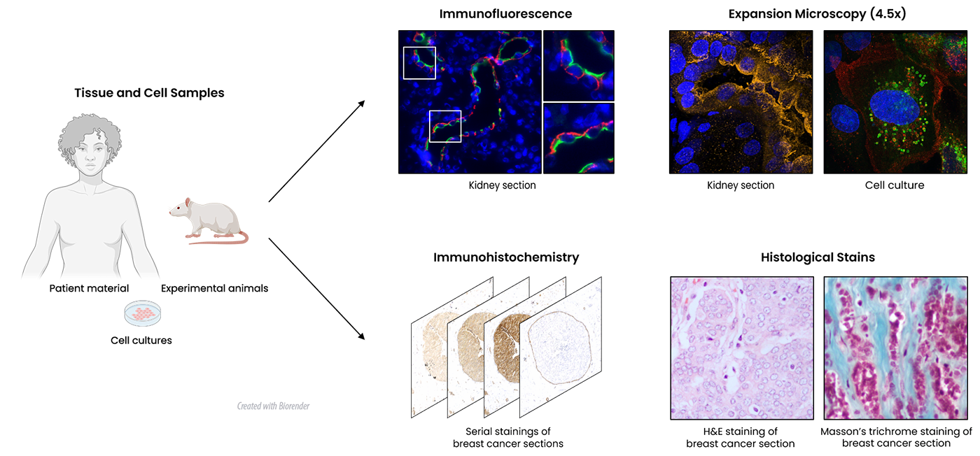
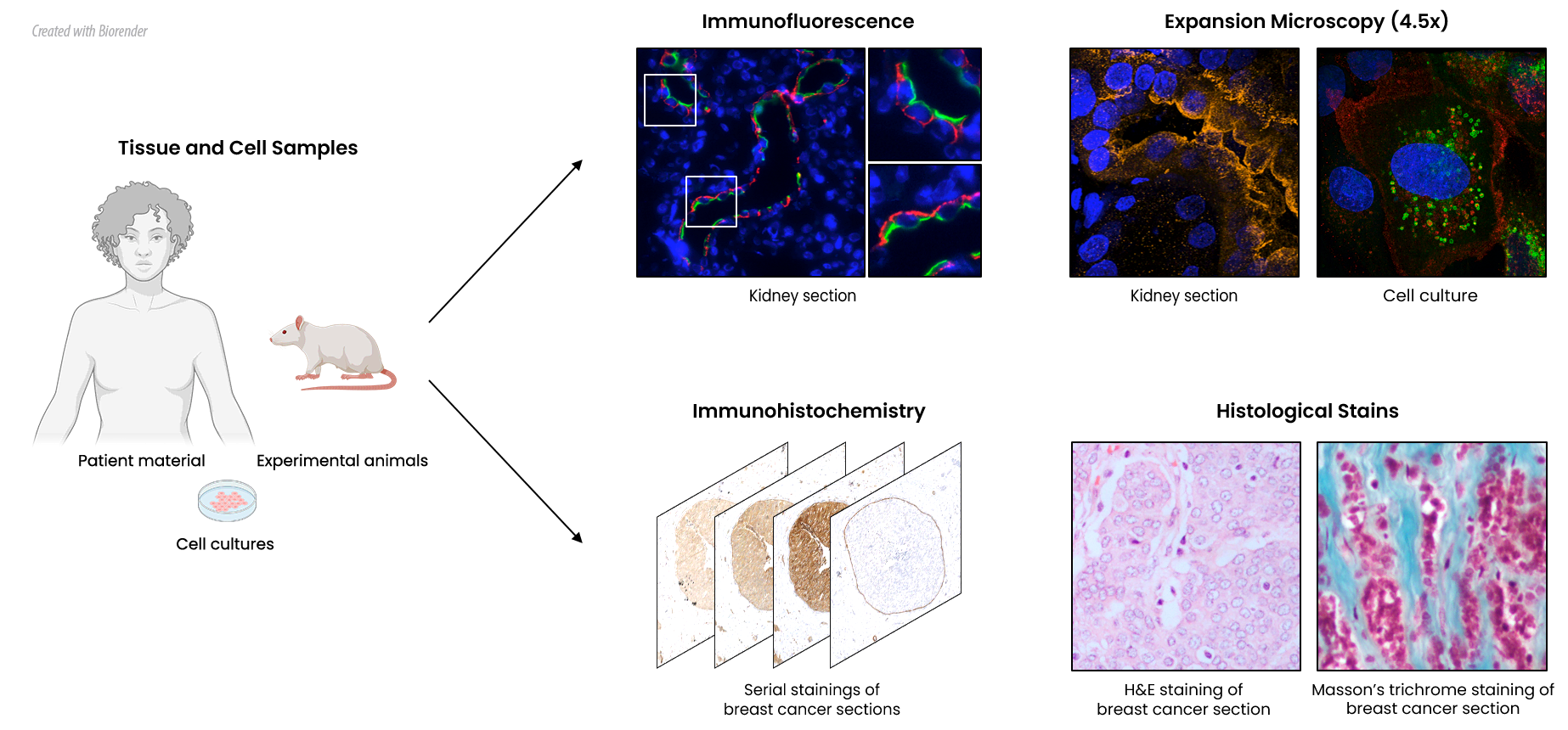
This figure illustrates the laboratory's expertise in bioimaging and sample analysis, highlighting key techniques such as fluorescence microscopy and Expansion Microscopy. It also showcases the use of immunohistochemistry and histological staining for the evaluation of patient-derived specimens.
Fluorescense Microscopy
Human Kidney
Multiplex; AQP1 (green, proximal tubules), AQP3 (yellow, collecting ducts), Uromodulin (magenta, thick ascending loops of Henle), Podocin (red, glomeruli), Prostaglandin E2 receptor 4 (white, glomeruli) and blue (nuclei)
Rat Kidney
AQP3 (yellow, renal collecting ducts) and nuclei (cyan)
Human Breast Cancer
AQP5 (magenta) and nuclei (cyan)
Mouse brain
AQP1 (red, choroid plexus) and nuclei (blue)
Expansion Microscopy & eSRRF
Rat kidney
AQP2 (magenta) original and post eSRRF processing
Renal Collecting Ducts
AQP2 (green, renal collecting ducts) and nuclei (blue)
Renal Collecting Ducts
AQP2 (green, renal collecting ducts)
MDCK Cells
AQP2 (green), Tubulin (Red), F-Actin (Magenta)
Time-lapse Microscopy
Migrating Cells (nuclei)
Wound healing assay; MDCK cells (nuclei in greyscale) and Velocity Analysis
Migrating Cells
Wound healing assay; EGFP (green) expressing MDCK cells
Zebrafish
Development (Vasculature)
Endocytic Trafficking